Computer Assisted for Prostate Brachytherapy Intervention
Coordinator (person and lab) Dimitris Visvikis / Julien Bert, LATIM
CAMI Partners: TIMC (30%), ISIR (10%), LTSI (20%), ICUBE (10%)
Started: January 2015 / 95% complete (1st recruitment: January 2015; expected closing date 12/2018)
The ambition of the CAPRI project is to provide an innovative brachytherapy treatment framework, less invasive, with fewer side effects, in rupture with current procedures, capable of accurately irradiating very localized areas within the prostate, while significantly decreasing the time of intervention.
Motivations and objectives:
Prostate cancer is by far the most common cancer in men. A large variety of prostate cancer treatments are available today in clinical routine. However, most of them act on the entire gland and are characterized by a high probability of side effects that considerably impact the patient’s quality of life. From an economic point of view, the high side-effect rates of these therapies lead to particularly expensive aftercare costs. The search for improved solutions in the treatment of prostate cancer remains a major societal challenge. This issue is emphasized by the fact that constant improvement of prostate cancer diagnostic tools allows detecting highly localized and small tumors at early age. In such cases, whole-gland treatment approaches are highly controversial. The high risk of side effects often justifies the choice of an active patient monitoring rather than therapy action. In recent years, a very attractive therapeutic alternative treatment concept is gaining in popularity among experts: focal therapy. This is a localized treatment, restricted to cancerous zones, with the objective of preserving healthy functional tissues inside and outside of the organ, and thus the quality of patient life. Finally, focal therapy has a high potential to reduce the intervention cost and duration, as well as the cost of aftercare.
However, in order to envisage in the future, the implementation of such focal treatment protocols using brachytherapy there is a need for current standard brachytherapy treatment procedures to fully encompass the latest state of the art in dosimetry, guidance and pre/per-operative imaging capabilities. Within this context, the ambition of the CAPRI project is to facilitate the development of an innovative brachytherapy protocol, less invasive, with fewer side effects, in rupture with current brachytherapy procedures, capable of accurately irradiating localized areas, while significantly decreasing overall intervention times.
In order to enable such an innovative brachytherapy framework, the CAPRI project is structured around different developments covering augmented perception, decision and finally action aspects.

Within the augmented perception context, new multi-modal image-fusion algorithms and processing (filtering, segmentation) for both pre-operative and per-operative tasks will be proposed. A specific challenge concerns the improvement of dosimetry accuracy. This calculation, already critical in global brachytherapy, is even more important when it comes to target small areas of the prostate.
We propose the development of the first ever clinical treatment planning system that integrates edema prostate model and a fast and personalized Monte Carlo dosimetry calculation on GPU.
Finally, haptic guidance will be proposed to the surgeon through the development of a brachytherapy application specific collaborative robot, in an attempt to improve treatment delivery (augmented action).
Main results and on-going work
- Augmented perception: enhancing US prostate image
A major limitation of Transrectal Ultrasound (TRUS) used per-operatively is the associated low image quality. TRUS images suffer from low contrast to noise ratio and weak prostate edges, rendering the prostate identification challenging. In this context, a new image enhancement methodology was proposed to facilitate further post-processing steps such as edge detection, US-MR co-registration or prostate segmentation. The proposed approach couples sharpening with anisotropic diffusion filtering within the partial differential equation (PDE) framework. The sharpening action is controlled by a new nonlinear hyperbolic shock filter term based on active contours external force fields in which edges are dynamically estimated and sharpened. Results show improvement with respect to state-of-the-art ultrasound denoising techniques such as Speckle Reducing Anisotropic Diffusion (SRAD) or an adaptive Voltera-type memory mechanism based on speckle statistics (ADMSS).
- Augmented perception: prostate segmentation
Segmentation of the prostate volume in TRUS images is generally performed in the clinic using tedious slice-by-slice manual delineations. We proposed a new quasi-automatic prostate volume segmentation method based on hybrid edge-region deformable models. More specifically, we combined statistical region forces based on the Bhattacharyya distance with a new prostate edge detector to better identify the prostate boundary. We also proposed a new quasi automatic initialization scheme by analyzing the radial edge profiles of the new edge detector. The approach only requires the selection of the apex and base slices, lowering the variability of results compared to manual initialization. The results show superior to state-of-the-art segmentation performance according to various segmentation metrics commonly reported in the literature. The proposed method was implemented within CamiTK.
- Augmented decision: fast and personalized dosimetry
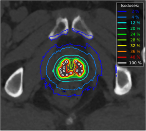
In this task, an accurate, personalized and fast Monte Carlo simulation (MCS) on graphics processing units (GPU) dedicated to brachytherapy applications was conceived. The aim was to estimate the dose deposition of the treatment within the patient as fast as possible without introducing any approximation compared to the real deposited dose. This simulation must consider organ heterogeneity, inter-radioactive seeds’ interactions and accurate source modeling. GGEMS is an advanced Monte Carlo simulations platform using GPU architecture. This software was extended and validated for brachytherapy showing high accuracy and fast calculation times. The proposed code allows a dosimetry calculation to be performed in 2 seconds considering a dose estimation uncertainty of 2% inside the prostate. In order to improve the accuracy of the simulation, a new hybrid particle navigator was developed in order to consider the true geometry of the 125I seeds (Source Tech Medical STM1251) within the patient.
- Augmented decision: prostate edema modeling
Prostate edema due to brachytherapy intervention has a negative effect in the outcome’s quality. Moreover, edema varies between patients and existing models are very general to be able to estimate accurately the patient-specific evolution of edema. We have therefore developed a biomechanical model where prostate is modeled as a biphasic material, composed of a fluid and a solid compartment. A Finite Elements method was used to simulate the edema formation by a linear increase in the pressure of the fluid, which generates the swelling of the solid. The amount of swelling is controlled from the patient-specific mechanical parameters of the prostate which can be estimated in-vivo (e.g. elastography). This allows providing a patient-specific solution for the prediction of edema.
- Augmented decision: advanced treatment planning system
The selection of seeds implantation sites is an arduous task and inverse planning is used to facilitate the procedure. Current treatment planning systems are based on the optimization of dose values recorded at sampled points over the patient’s anatomy which do not correspond to the deposited dose in the volume of the organs. To address this limitation, we developed an inverse planning method where the dose received from the whole volume of the organ is optimized. In each iteration of the optimization method the dose volume histogram (DVH) is extracted and optimized using Fast Simulated Annealing. This allows delivering high and homogeneous dose to the prostate while reducing the dose deposited to the organs at risk, compared to current clinical planning systems.
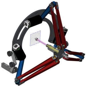
- Augmented action: haptic guidance with collaborative robot
To enhance the precision of the insertion of the needles in the prostate, given a target position provided by the treatment planning system, a collaborative robot dedicated to brachytherapy has been proposed. The aim is to let the surgeon control the needle insertion with a haptic guidance provided by the cobot. Direct and inverse kinematic models were estimated. In addition, we developed an advanced haptic control (hardware and software) based on a force impedance control system. A first prototype was manufactured and is currently undergoing performance evaluation.
Transfer : 25% clinical / 0% industrial transfer
-
Retrospective analysis on over 200 patients undergone whole gland low dose brachytherapy to assess the correlation of dose (calculated based on standard dosimetry tools) with organ toxicity (promoter: CHRU Brest; starting date 10/2017)
-
Phase 1 clinical trial evaluating the interest of elastography TRUS for the determination of patient specific prostate elasticity parameters within the context of dosimetry in prostate brachytherapy (promoter CHRU Brest; starting date 12/2017)
-
Retrospective analysis of multiparametric MRI and PET imaging for the definition of target volumes in prostate brachytherapy (CHRU Brest, CRLCC Rennes, CHU Poitiers, APHP, starting date 1er trimester 2018)
Most recent publications on the project
-
Jaouen and L. Gaubert and J. Bert and M. Hatt and D. Visvikis. Image Filtering with Advectors. IEEE International Conference on Image Processing, 2018.
-
Jaouen, J. Bert, N. Boussion, H. Fayad, M. Hatt, D. Visvikis. Image enhancement with PDEs and nonconservative advection flow fields. Transactions on Image Processing, 2017 (in press)
-
Jaouen V, J. Bert, I. Hamdan, A. Valeri, U. Schick, N. Boussion, D. Visvikis. Purely edge-based prostate segmentation in 3D TRUS images using deformable models. SURGETICA, 2017.
-
Bert J, Y. Lemaréchal, D. Visvikis D. New hybrid voxelized/analytical primitive in Monte Carlo simulations for medical applications. Physics in medicine and biology, 61 pp 3347-3364, 2016.
-
Mountris, J. Bert, D. Visvikis. Edema adapted treatment plan in LDR prostate brachytherapy: beyond common inverse treatment planning. European Society for Radiotherapy & Oncology (ESTRO), 2018.
-
Mountris, J. Bert, J. Noailly, A. Rodriguez Aguilera, A. Valeri, O. Pradier, U. Schick, E. Promayon, M. A. Gonzalez Ballester, J. Troccaz, D. Visvikis. Modeling the impact of prostate edema on LDR brachytherapy: a Monte Carlo dosimetry study based on a 3D biphasic finite element biomechanical model. Physics in Medicine and Biology, 62 pp 2087—2102, 2017.
-
Mountris, D. Visvikis, J. Bert. DVH-based Inverse Planning using Monte Carlo Dosimetry for LDR Prostate Brachytherapy. International Journal of Radiation Oncology*Biology*Physics, 2018 (in press).
-
Mountris, J. Bert, D. Visvikis. LDR prostate brachytherapy inverse planning including dose-volume relation and tissue heterogeneity. European Society for Radiotherapy & Oncology (ESTRO), 2018.
CAPRI Team:
Project Managers
Julien Bert
PostDocs
Vincent Jaouen
Iyas Hamdan
Konstantinos Mountris
PhD student
![]()
![]()
![]()
1 thought on “CAPRI”
Comments are closed.